
Symptoms, medical history and examination of the teeth and tissues
Seeing what the examination alone does not show
Dental diagnosis and radiology
Targeted examinations to understand the situation, gauge the risks and build a proportionate treatment plan.
- Recovery
- No anaesthesia or recovery needed
- Results
- Outpatient examination, no hospital stay
Price and stay
Dental Diagnosis and Radiology
FreeBase price
- Hotel
- Hotel not includedFor treatments over €10,000, one week of hotel accommodation is complimentary on the first stay.
Included
- panoramic X-ray
- clinical examination
Base price shown as a guide: your personalised quote, drawn up after assessment, is the one that applies.
Get a personalized quoteGoals and benefits
Dental diagnosis and radiology
Dental radiology complements the clinical examination when information cannot be obtained by observation alone.
The benefits
- Examination chosen according to the clinical question
- Images stored in the record and interpreted together with the clinical assessment
- 2D or 3D visualisation adapted to the area studied
- Support for implant and surgical planning when needed
- Detection of certain conditions not visible on clinical examination
- Protocols aimed at limiting radiation exposure
- Recent usable examinations can be reused and shared
- Targeted follow-up according to the progress of the treatment
Choosing the right examination for the right question
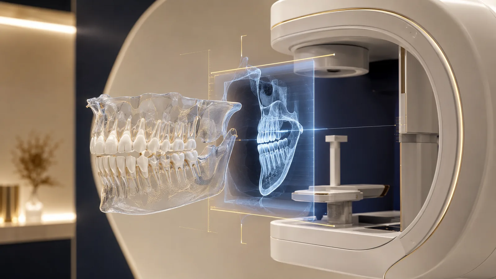
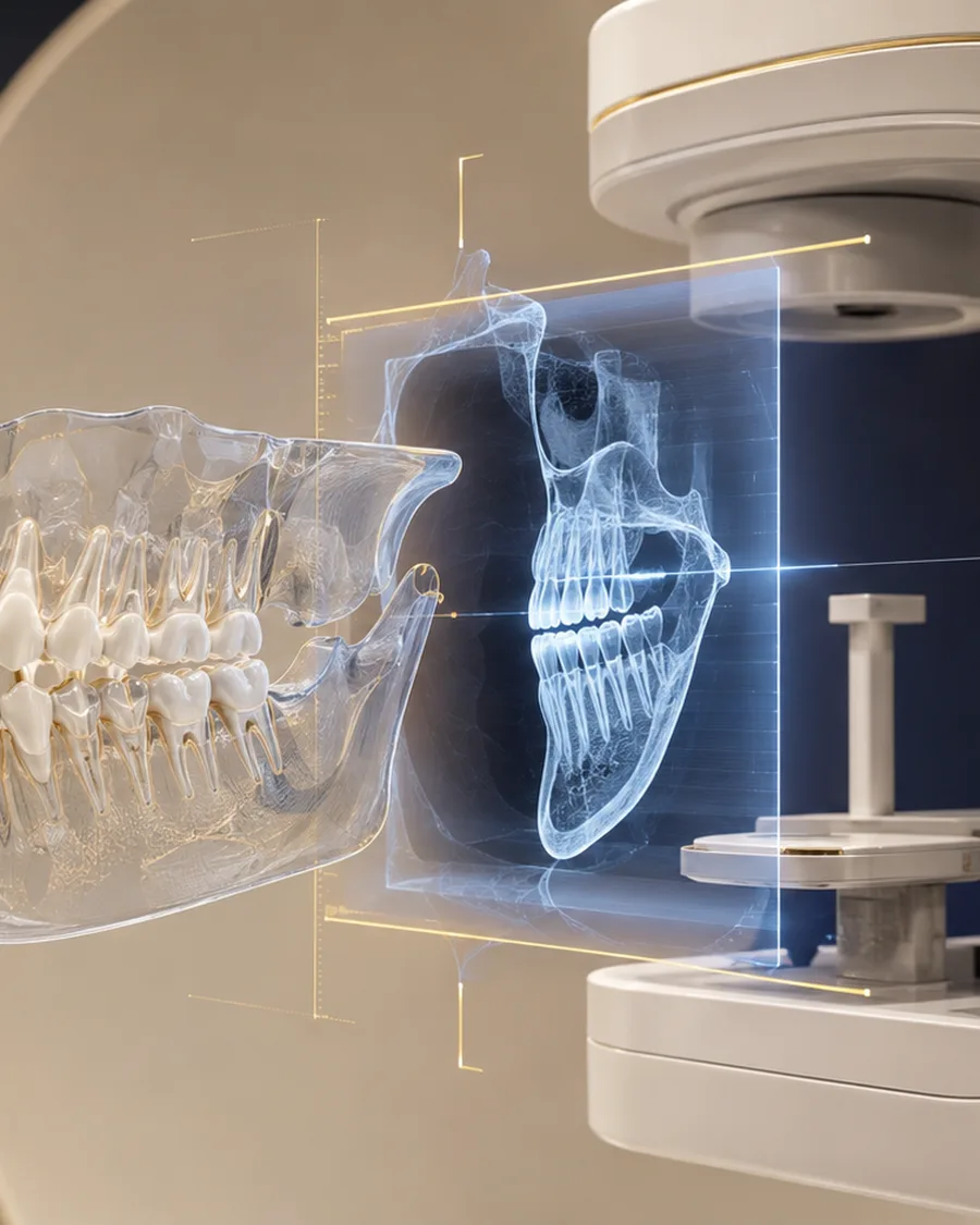
An intraoral radiograph, a panoramic radiograph and a cone beam scan do not provide the same information. The examination is chosen according to the clinical question, while limiting unnecessary exposure.
- Examination justified by a clinical question
- Interpretation combined with the symptoms and the examination
- Data used to plan and to monitor
What the practitioner assesses
Area to be explored and level of detail required
Previous examinations available and expected benefit
Treatment video
Educational animation of the main treatment stages. The actual protocol is adapted to each patient's diagnosis.
Procedure
The treatment process
Clinical examination and diagnostic question
The practitioner records symptoms, history and previous treatments, and examines the teeth, gums, occlusion and tissues. They then determine whether an image is necessary and which technique is the most suitable.
Choice of the appropriate examination
A targeted X-ray, a panoramic or a cone beam is selected according to the area and the clinical question. The 3D scan is chosen only when it provides useful information that simpler imaging does not sufficiently supply.
Image acquisition
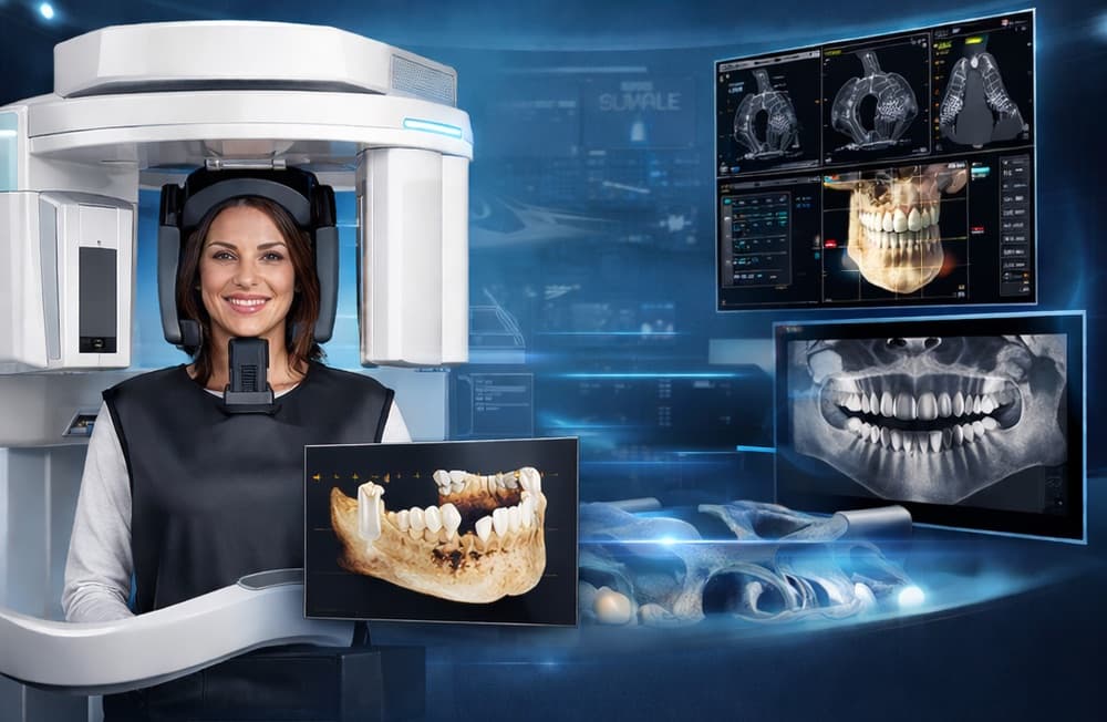
The patient is correctly positioned and must remain still for a few seconds. For a cone beam, the device rotates around the head to collect the data needed for the 3D reconstruction.
Interpretation and planning
The images are analysed together with the clinical examination to identify structures, anomalies and treatment constraints. An additional interpretation may be requested when an area goes beyond the practitioner's usual field.
Storage, sharing and targeted follow-up
The images and conclusions are kept in the record. They can be shared with the patient or the professionals concerned. A new examination is performed only if it provides additional clinical information.
Technology and materials
Digital radiography and cone beam
The settings and the field of view are adapted to the indication. Images must be interpreted in their clinical context.
Safety
Contraindications
- Radiological examination prescribed only when it can change the diagnosis or the treatment
- Choice between a 2D image and cone beam according to the clinical question, without systematic 3D scanning
Frequently asked questions
Is a cone beam scan necessary for every treatment?
No. It is prescribed when the 3D information is useful and justified. A simpler radiograph may be sufficient.
Can I send my previous radiographs?
Yes. Recent, good-quality examinations can avoid some repeat imaging, provided that they answer the clinical question.
Related treatments
Request your quote
Ready to transform your smile?
Send us your panoramic X-rays and photos to receive a free assessment and a detailed treatment plan within 24 hours.
Free consultation within 24h
Updated on