
Reason for the extraction and prognosis of conservative treatments
Removing a tooth that cannot be saved
Tooth extraction
An extraction is proposed when a tooth cannot reasonably be saved or when its presence creates a risk for the neighbouring tissues.
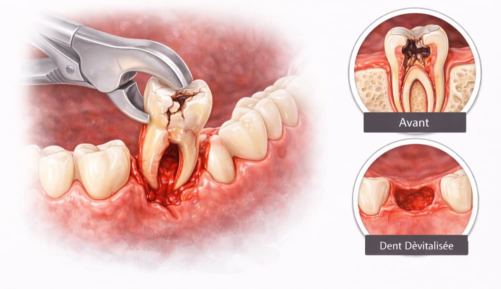
- Recovery
- Discomfort, swelling and sensitivity possible for several days
Price and stay
Tooth Extraction
€10Base price
- Hotel
- Hotel not includedFor treatments over €10,000, one week of hotel accommodation is complimentary on the first stay.
Included
- anaesthesia
- post-operative follow-up
Not included
- flights
- medication
- hotel
- transfers
Base price shown as a guide: your personalised quote, drawn up after assessment, is the one that applies.
Get a personalized quoteGoals and benefits
Tooth extraction
A tooth extraction consists of removing a tooth when it cannot be preserved predictably, or when keeping it carries more risks than benefits.
The benefits
- Removal of a non-preservable tooth under local anaesthesia
- Simple or surgical extraction chosen according to the anatomy
- Planning that takes the roots, nerves and sinuses into account
- Bone preservation or a graft discussed according to the future prosthetic project
- Instructions and post-operative check-up adapted to the difficulty of the extraction
- Immediate implant possible only in selected cases
- Possibility of planning a bridge, denture or implant after healing
- Management of dry socket or infection if it occurs
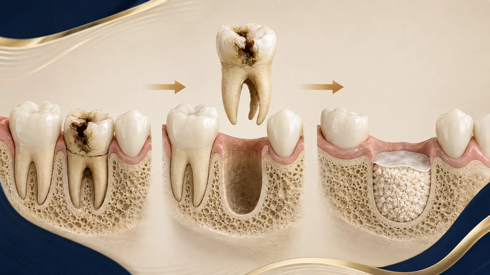
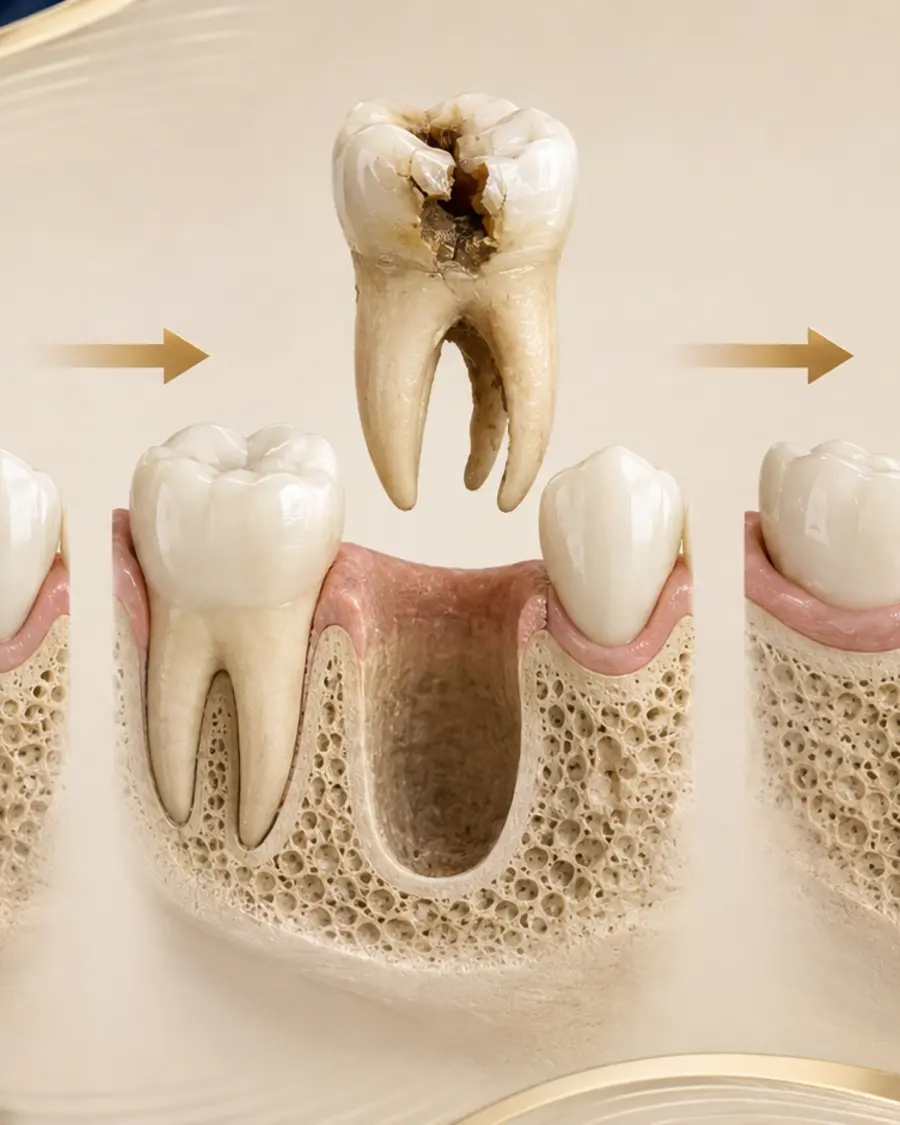
Preserving as much bone and tissue as possible
The technique depends on the shape of the roots, the position of the tooth and the surrounding anatomy. The replacement plan should be discussed before the extraction whenever this is possible.
- Prior check of the options for saving the tooth
- Technique adapted to the anatomy of the tooth
- Planning of the replacement and of the healing
What the practitioner assesses
Shape of the roots and proximity of nerves or sinuses
Infection, medication, blood clotting and prosthetic plan
Treatment video
Educational animation of the main treatment stages. The actual protocol is adapted to each patient's diagnosis.
Procedure
The treatment process
Diagnosis and possibility of preserving the tooth
Clinical and radiographic examination to confirm that the tooth cannot be preserved predictably, evaluate the roots, the bone and the neighbouring structures, then plan the possible replacement.
Anaesthesia and medical preparation
Local anaesthesia is administered. Medical treatments, allergies and medications affecting bleeding or healing are checked before the procedure.
Simple or surgical extraction
The tooth is mobilised and removed. Depending on its position and shape, an incision, sectioning of the tooth or limited bone removal can be necessary.
Socket cleaning and bleeding control
The socket is inspected and rinsed when necessary. Compression, a haemostatic material or sutures can be used. A preservation graft is discussed according to the future project.
Instructions, healing and follow-up
The instructions aim to protect the clot and limit complications. A check-up is organised according to the difficulty. Increasing pain after a few days, a bad taste, fever or persistent bleeding must be reported.
Technology and materials
Targeted imaging and socket preservation
An X-ray or a cone beam scan may be indicated. Depending on the plan, a biomaterial can help to preserve the volume, without guaranteeing the absence of bone remodelling.
Safety
Contraindications
- Anticoagulants, antiplatelet drugs or clotting disorders must be reported; no treatment should be stopped without medical advice
Frequently asked questions
When can the extracted tooth be replaced?
The replacement can be immediate or delayed depending on the infection, the bone, the tissues and the stability that can be achieved. The choice is made before or at the time of the procedure.
Which signs should lead to a consultation after an extraction?
Persistent bleeding, increasing pain, a fever, significant swelling or difficulty swallowing require you to contact the clinic.
Related treatments
Request your quote
Ready to transform your smile?
Send us your panoramic X-rays and photos to receive a free assessment and a detailed treatment plan within 24 hours.
Free consultation within 24h
Updated on